
How to achieve an optimal image
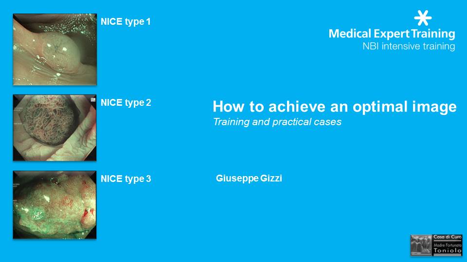
Why is it so important to achieve, analyze and store high quality images in digestive endoscopy? …simply because Digestive Endoscopy is a discipline based on images and pictures, and therefore the quality of the images is the cornerstone in order to DETECT, CHARACTERIZE and CLASSIFY an early neoplastic lesions, particularly in the colorectum.
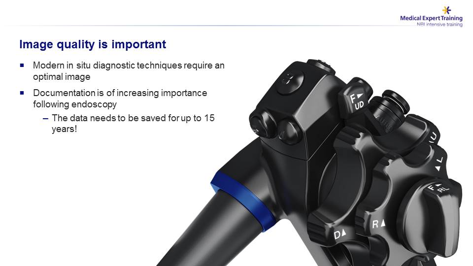
Also the possibility to file or store relevant images is an important endowment for the endoscopist; it allows to compare the endoscopic appearance with the histology report and, hence, verify ability and accuracy of the diagnostic prediction.
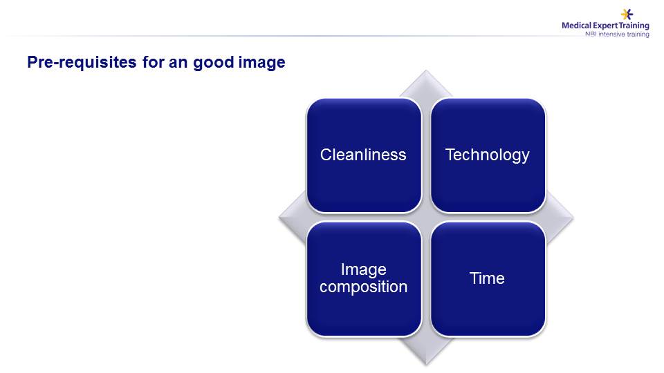
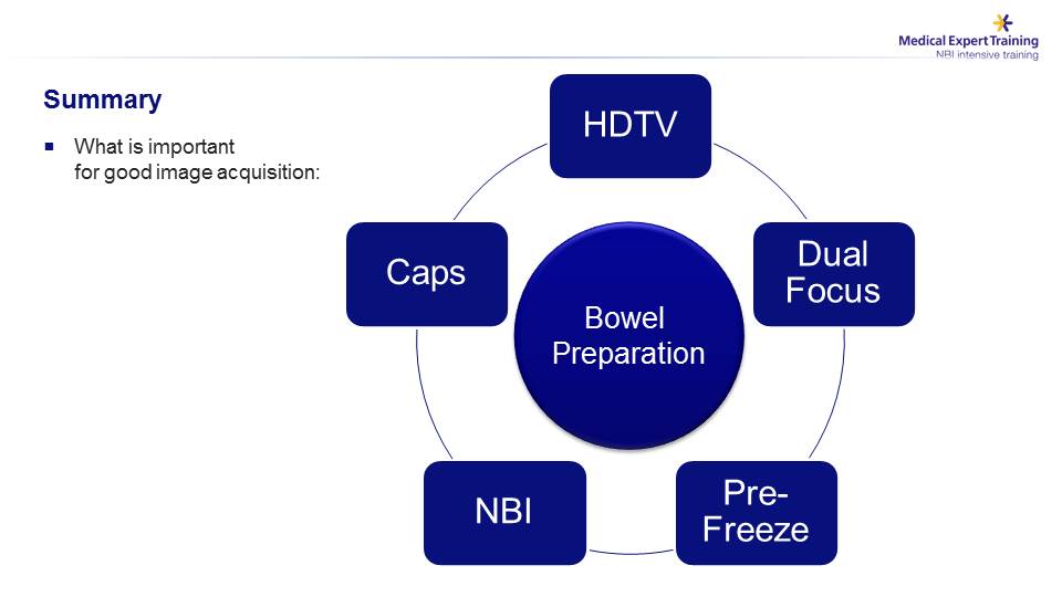
Preconditions to obtain high quality endoscopic images are: a) adequate bowel cleansing; b) advanced endoscopic equipment and technology; c) ability to correctly focus the lesion and; last but not least, d) the possibility and willingness to be committed with the necessary time to this specific goal.
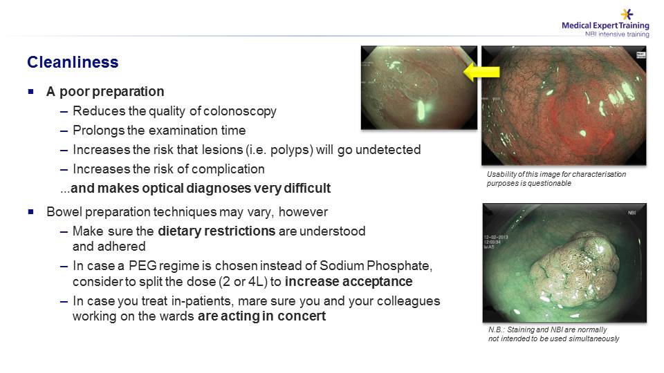
It is unnecessary to remind that an inadequate or poor bowel cleansing not only does jeopardize any detailed inspection of the lesion, but it can even completely mask the lesion to the eye of the endoscopist…..
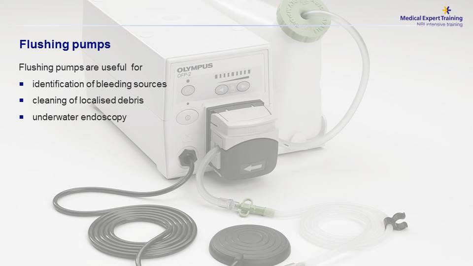
…. and therefore it is important to have a flushing water pump to wash away any fecal residue and also obtain the so called “immersion view”, sometimes extremely useful for a closer assessment of the lesion….
… this video shows the usefulness of the flushing water pike pump in the assessment of a serrated lesion of the ascending colon covered by the typical “mucus cap” .
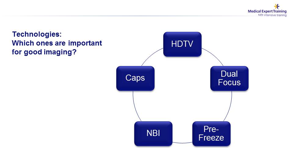
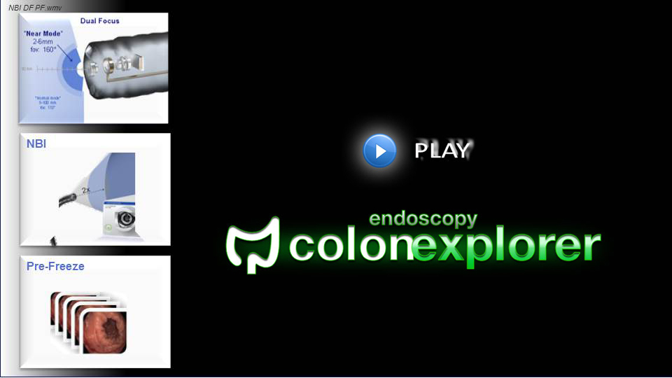
As far as the technological aspects are concerned, the necessary parameters to achieve high-quality diagnostic images are: 1) the use of a distal transparent CAP; 2) the availability of a FULL HD resolution equipment; 3) the Dual Focus technology; 4) the NBI technology; 5) the Pre-Freeze function.
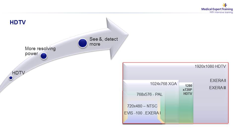
Higher resolution allows an increased capability to visualize and assess the lesions…..
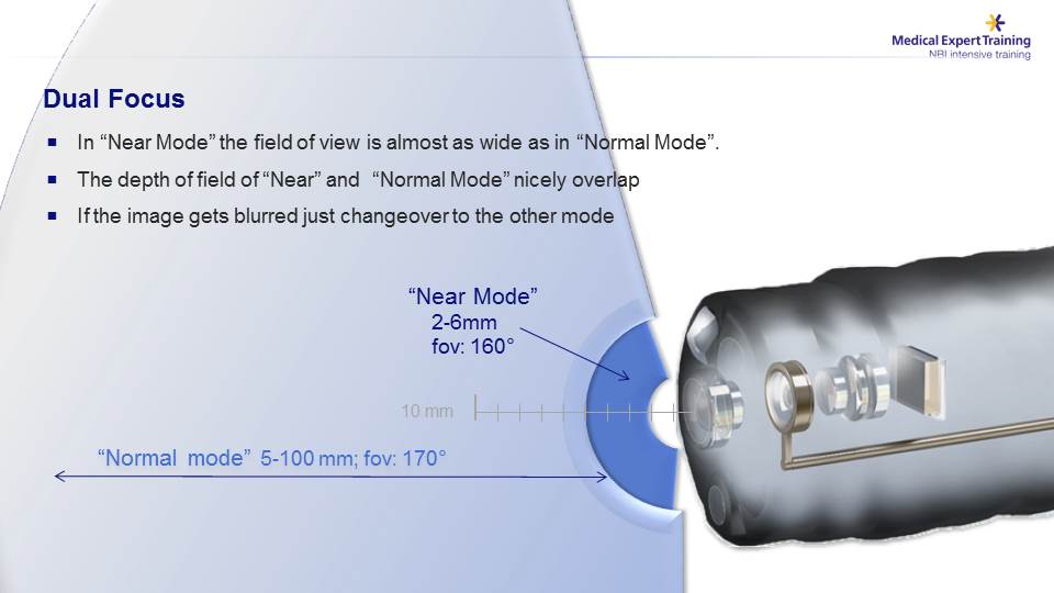
The DUAL-FOCUS technology offers the chance to select both a panoramic view (“Normal mode”), employed during the standard endoscopic exploration of the mucosa, or a closer view (“Near mode”), when the endoscopist proceeds to the CHARACTERIZATION of the lesion….
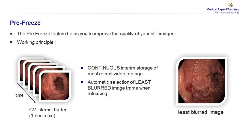
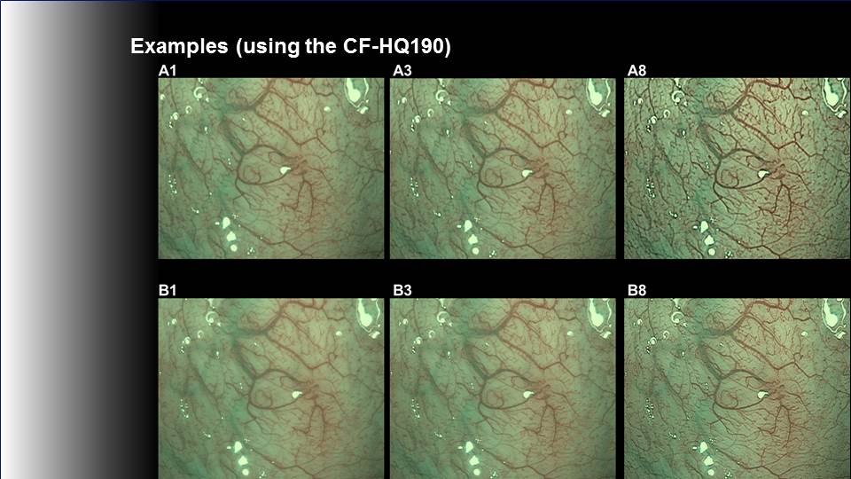
By using the PRE-FREEZE function, the EXERA III system automatically selects from a buffer (a sort of continuously updated image source), the most clear image which can then be archived.
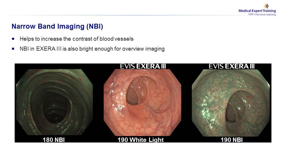
Another high-tech function is the NBI, that allows to enhance the mucosal and submucosal capillary vessels , as shown in the slide… The Olympus Exera III system provides for the partial loss of brightness reported with the 180 series, enabling an excellent visualization with a depth of field equivalent to that of the white light
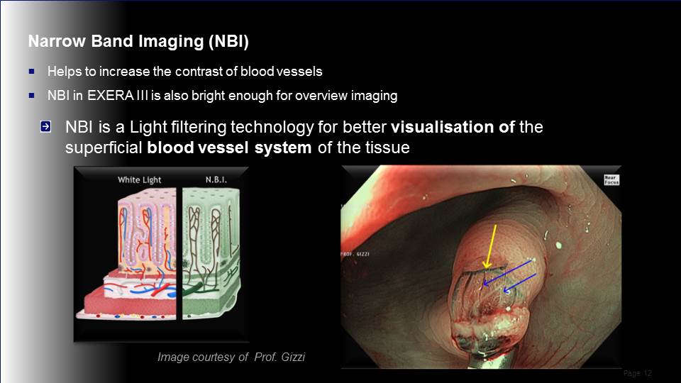
……the most superficial vessels, i.e. intramucosal capillary vessels show a brownish color (blue arrows), while the deeper vessels show a green color (yellow arrow)….
…… this video summarizes the practical use of these three functions: Dual Focus, NBI, Pre-Freeze, and underscores their extreme usefulness in the characterization of a lesion in closer view, in this case a small adenomatous polyp ….
The NBI function can be modulated specifically for the capillary vessels ….
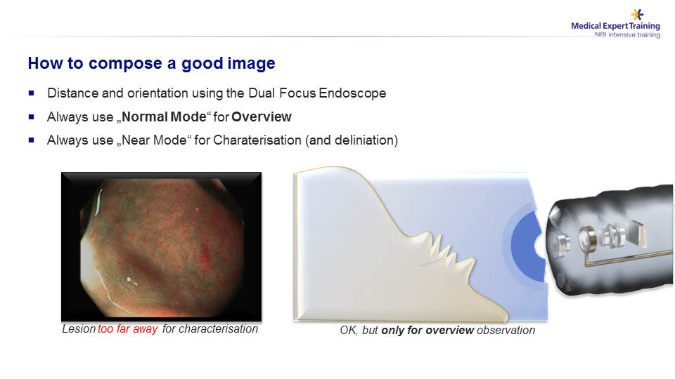
To correctly assemble the endoscopic image it is crucial to position the distal tip of the scope perpendicular to the lesion, rather than tangentially to avoid “out of focus” areas ….
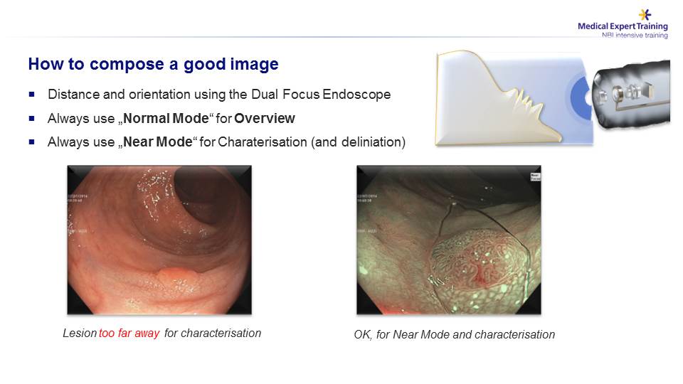
After the identification of the lesion (image in white light) the “Near Mode” view, combined with the NBI facilitate the characterization of the lesion.
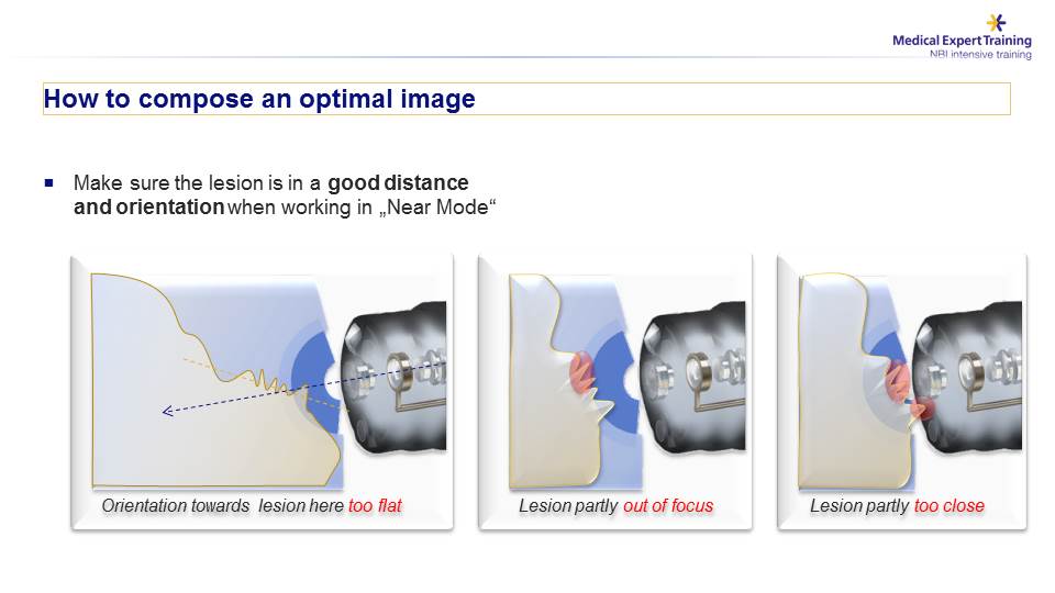
The endoscopic view with the “Near mode” modality imply the correct focal distance and coherent orientation with the direction of the light beams

The following sequence shows how to achieve the right orientation and distance in relation to the lesion.
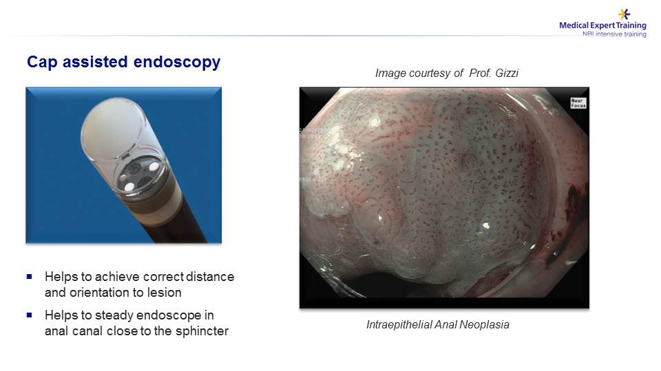
In some GI segments the cap attached onto the tip of the scope is an essential device to keep focus on the lesion, especially in areas adjacent to sphincteric structures.
In other circumstances, the diathermic snare may be used to make the area to evaluate stable (in the video the assessment of the resection margins after a mucosectomy).
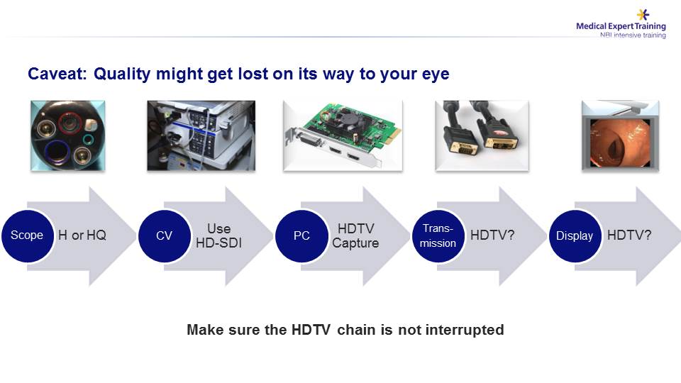
It is obvious, though, that high quality images captured by the endoscope and elaborated by the video-processor requires that all the further transmission steps toward the monitor or the capture graphic board are achieved with no loss of definition.
Achieving high quality images in digestive endoscopy means the possibility to guarantee to our patients an extremely accurate diagnostic procedure This can be obtained only by employing advanced technology, in the hands of an expert endoscopist who can devote adequate time to this complex investigation.
The key to quality endoscopic examinations is the commitment of adequate time to take advantage of the resources provided by high-tech endoscopic equipment